Caustics and Corrosives - Medical Toxicology Guide
Introduction
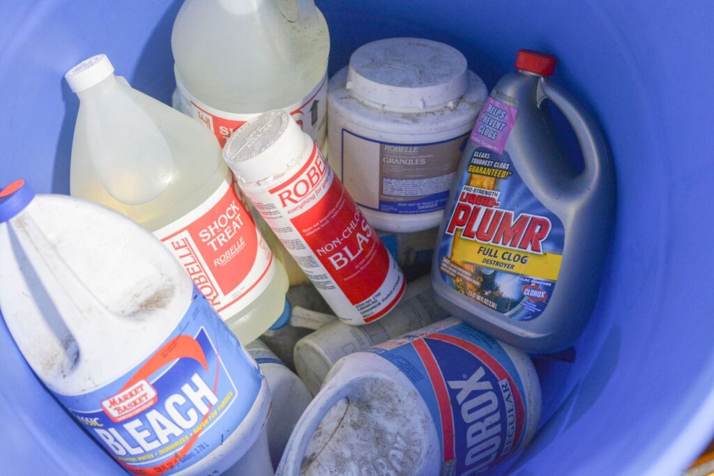
Caustics and corrosives result in tissue injury by a chemical reaction. The majority of caustic substances are either acidic or alkaline. The degree of tissue injury from acidic and alkaline substances is influenced by several factors such as the duration of contact with the tissues, the amount and state (liquid, solid) of the substance involved. Other physical factors include the pH, concentration, and the ability to penetrate the tissues. Alkaline drain cleaners and acidic toilet bowl cleaners are responsible for the most fatalities from corrosive agents.
Toxic Dose
Variable: The toxic dose varies tremendously by type and concentration of acid.
Epidemiology
- Poisoning is common.
- Toxic effects following exposure are typically mild.
- Mortal effects (deaths) occur after ingestion of a large amount of dilute solution or a smaller amount of concentrated, highly acidic/alkaline compounds.
- Occupational exposure to acid/alkali mists is associated with laryngeal cancer.
Causes
- Toxic ingestion is usually accidental in children.
- Adult toxicity is most likely to result from suicidal or occupational exposure.
Mechanism of Toxicity
- Caustics are typically classified as acids or alkalis. An acid is a proton donor and causes significant tissue injury when the pH < 3. An alkali is a proton recipient and causes significant tissue injury when the pH > 11.
- Acids cause coagulative necrosis. Hydrogen ions desiccate epithelial cells, causing edema, erythema, tissue sloughing, and necrosis, with the formation of ulcers and eschars.
- Alkalis cause liquefactive necrosis, a process that involves saponification of fats and protein dissolution. Cell death occurs as a result of emulsification and disruption of cellular membranes. The hydroxide ion in alkaline agents reacts with tissue collagen, causing it to swell and shorten. Small vessel thrombosis and heat production occur.
Clinical Manifestations
- Signs and symptoms of impending airway irritation or obstruction: Dyspnea, stridor, hoarseness, dysphonia or aphonia, respiratory distress, tachypnea, hyperpnea, cough, and chest pain.
- Other symptoms and signs of injury: Nausea, vomiting, oropharyngeal burns (pain or swelling). Significant esophageal damage may occur even in the absence of oropharyngeal lesions. Other symptoms include drooling, dysphagia, abdominal pain, subcutaneous air, hematemesis, melena, tachycardia, acute peritonitis (abdominal guarding), rebound tenderness, and diminished bowel sounds.
- Signs of severe injury: Altered mental status, peritoneal signs, perforation, hypotension, stridor, renal failure, electrolyte disturbance, metabolic acidosis, hemolysis, and shock.
Differential Diagnosis
- Chemical Burns
- Thermal Burns
- Dysphagia
- Gastrointestinal hemorrhage or perforated viscus
Injury Severity and Complications
| Grade | Findings | Comments |
|---|---|---|
| Grade 0 | No injury | No adverse effects |
| Grade I | Erythema, edema | No adverse sequelae |
| Grade IIa | Limited to mucosa | No adverse sequelae |
| Grade IIb | Blister, ulceration, and whitish membrane penetrating the mucosa | May develop strictures |
| Grade III | Extensive necrosis, thickness, or frank perforation | High morbidity and mortality may result in perforation, mediastinitis, or peritonitis. Strictures are seen in the majority. |
Investigations
Routine
- Bedside RBS
- ABGs, Electrolytes
- CBC, BUN, Creatinine, ALT, AST, Creatine Kinase
- EKG
- Pregnancy test for females of childbearing age
Specific
- Determine type and cross-match for blood, and monitor urine output.
- Serum lactate and base deficit are useful prognostic tools.
- MetHb levels, LDH for (phenol), calcium for (oxalates, fluorides), and urine oxalate.
- Chest X-ray to evaluate for pneumomediastinum or free air under the diaphragm. The absence of these findings does NOT rule out the possibility of necrosis or perforation of the esophagus or stomach.
- Abdominal X-ray to evaluate for pneumoperitoneum, ascites, or an ingested button battery (metallic foreign body).
- CT scan is useful in detecting small amounts of extraluminal air not caught on plain radiographs.
- Several weeks after ingestion, barium contrast X-rays of the upper GI tract are useful in patients with grade II or III burns to evaluate for strictures.
Treatment
Stabilization
- Airway, breathing, and circulation (ABC) should be evaluated and stabilized as necessary.
- Nothing per oral (NPO) and IV fluids until complete evaluation.
- Do not force the patient to vomit or drink.
- Allow the patient to drink a small amount of water if able to check for swallowing ability.
Decontamination
- Ocular burn: Rinse with 0.9% sodium chloride or running tap water at 30-32°C for 15 minutes, followed by emergency ophthalmologic consultation.
- Dermal burn: Remove contaminated clothes, brush off particulate corrosives, and follow with copious irrigation.
Specific: Endoscopy
- Perform as soon as possible (best within 12 hours, not more than 24 hours) in any patient who ingested high volumes of a caustic substance. The grade of mucosal injury at endoscopy is the strongest predictive factor for complications and mortality. The absence of visible oral burns and edema does NOT reliably exclude esophageal or gastric burns.
- In patients with extensive circumferential burns (grade IIB or III), a nasogastric tube (NGT) should be placed under direct visualization during the endoscopic procedure. A NGT should not be inserted blindly to avoid perforation or additional injury. The NGT can provide nutritional support during the healing phase and help maintain a lumen during stricture formation. Consider radiological control 30 days after ingestion and an endoscopy 4-6 weeks after ingestion.
- Consult an otolaryngologist if stridor does not improve for possible tracheostomy.
- Consult a surgeon and perform abdominal X-ray & amylase if the patient experiences rebound tenderness, rigidity, or severe abdominal pain.
Supportive Treatment
- Analgesia: Paracetamol, NSAIDs, and morphine are permitted initially. H2 blockers or proton pump inhibitors IV infusion (0.7-3.5 mg/kg/day).
- Bronchospasm: Treat with oxygen, inhaled β agonists, and consider systemic corticosteroids. Consider mechanical ventilation if ventilation is compromised.
- Blood transfusion: If Hb is less than 8 gm/dl or rapid hemorrhage reduces Hb to <80% of expected value.
- IV fluid: Followed by total parenteral nutrition (TPN) if stage II or III corrosion, severe dysphagia, or respiratory compromise (inability to cough). Refer to TPN protocol.
- Antibiotics: Use when indicated.
- Steroids: The use of corticosteroids to prevent stricture formation is controversial.
- Oral fluids: Liquid diet may be permitted with caution if endoscopy was not performed and the patient does not complain of dysphagia or respiratory complications. Stop the oral diet if vomiting or bleeding occurs.
- Observe for evidence of bleeding (e.g., hematemesis, melena), nutritional status, and respiratory function.
- Observe for broncho-esophageal fistula that can form on the 7th-10th day by imaging (cough on drinking).
- Refer to the surgery department for treatment and follow-up of any possible complications from caustic injury.
Admission Criteria
- Symptomatic patients and those with endoscopically confirmed grade II or higher burns should be admitted.
- Patients presenting with respiratory distress, grade III burns, extensive grade II burns, metabolic acidosis, hemodynamic instability, gastrointestinal bleeding, or large ingestions should be admitted to an intensive care setting.
Common Pitfalls
- Diluting the corrosive by excessive drinking of water or milk as they may induce vomiting.
- Neutralizing the corrosive.
- Administration of syrup ipecac and performing gastric lavage.
- The use of activated charcoal as it is not effective and potentially hazardous.
- Insertion of Ryle tube blindly unless endoscopically guided.
- Blind naso or endotracheal intubation.
- The absence of oral burns does NOT reliably exclude the possibility of significant esophageal burns.
- Patients may have severe tissue necrosis and impending perforation requiring early surgical intervention without having severe hypotension, rigid abdomen, or radiographic evidence of intraperitoneal air.
- Patients with any evidence of upper airway involvement require early airway management before airway edema progresses.
- The extent of eye injury (degree of corneal opacity) may not be apparent for 48-72 hours after the burn.